
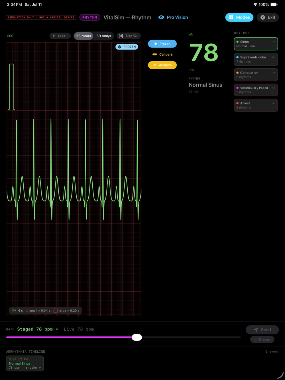
Ventricular tachycardia (VT)
A run of fast, wide QRS complexes from the ventricles — often regular and organized. It can be with or without a pulse; the patient, not the monitor, tells you which.
Ventricular fibrillation (VF)
Chaotic, disorganized electrical activity with no organized QRS — just a quivering baseline. It's the classic "shockable" arrest rhythm and the deadliest pattern on the monitor.
The arrest rhythms — and the pulse rule
Two more rhythms define cardiac arrest: pulseless electrical activity (PEA), where the monitor shows organized-looking electrical activity but there is no pulse, and asystole, the flat line. The lesson underneath them: electrical activity does not prove mechanical activity — always confirm the patient.
Recognize, then escalate
Your job with these rhythms is fast recognition and escalation — call for help and follow your team's and your facility's ACLS protocols. This guide (and the app) build the recognition; the response belongs to you, your team, and your protocols.
Practice spotting them — safely
VitalSim Rhythm lets you load VT, VF, and the arrest rhythms on a simulated strip and study their morphology as many times as you need — a safe place to build the pattern recognition you never get to rehearse on a real patient.

FAQ
What's the difference between VT and VF?
Ventricular tachycardia is fast, wide, and usually organized; ventricular fibrillation is chaotic with no organized QRS. VF is the classic shockable arrest rhythm.
Which rhythms are shockable?
Ventricular fibrillation and pulseless ventricular tachycardia are the shockable arrest rhythms; PEA and asystole are not. Always follow your ACLS and facility protocols.
What is PEA?
Pulseless electrical activity — organized-looking electrical activity on the monitor with no pulse. It's a reminder that what's on the screen doesn't prove there's a pulse.